The Nobel Prize in Chemistry this year was awarded to Eric Bettsigu, Stefan Helle and William Merner for the development of high-resolution fluorescence microscopy. Their names have not been mentioned in the past analysts’ forecasts, but well-deserved award and a great value of their work are undeniable.
Scientists who contributed to the development of microscopic techniques, not the first time attention is drawn to the Nobel Committee. In 1925 received the Prize in Chemistry Richard Adolf Zsigmondy for the study of colloidal solutions. One of the main achievements of Zsigmondy who played an important role in his discoveries, and in the further development of colloid chemistry, was the invention of ultramicroscope and immersion microscope. In 1953, the Prize, this time in the field of physics, received Dutchman Frits Zernike, the inventor of a phase contrast microscope. In 1982, Aaron Klug for the development of crystallographic electron microscopy method received the prize in chemistry. Particularly successful for the inventors of new types of microscopes appeared in 1986. Then Prize in Physics received Ernst Ruska – creator of the first electron microscope, and Gerd Binnig and Heinrich Rohrer – inventors of the scanning tunneling microscope. In the same year, but in the field of chemistry, received the Nobel Prize Aaron
Creating new microscopes, researchers tend to see smaller and smaller objects, and very soon on their way stands a barrier that seems insurmountable. This diffraction limit, or limit Abbe, named in honor of the German optics Ernst Abbe, who discovered this phenomenon in 1873. Abbe found that the minimum spot size that we can get, focusing electromagnetic radiation is given by λ / 2n, where λ – wavelength of the electromagnetic wave in a vacuum, n – refractive index of the medium. Microscope does not help us to see objects that are smaller than this value. The wavelength of the electrons is less than that of photons, so the possibility of electron microscopes more, but before them inexorably rises diffraction limit.
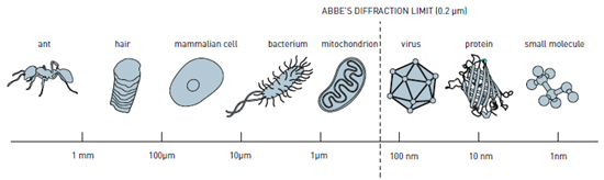
The diffraction limit (Abbe limit). The fact that the right border, in the microscope not be seen.
We once told how the Abbe limit is overcome by using a fundamentally different method – atomic force microscopy, no relation to the optics are not having. Today’s laureates have addressed this issue differently.
All three scientists working in the field of fluorescence microscopy based on the luminescence of the atoms and molecules of a sample in response to the absorption of radiation. In the fluorescence microscope the object is illuminated with light of a specific wavelength that is absorbed by the molecules, causing them to emit light of longer wavelengths (ie, a different color than the absorbed). This light is detected by the detector. By itself, the method of fluorescence microscopy is not able to overcome the Abbe limit.
In 1993, Stefan Kjell, who worked at that time in Finland, at the University of Turku, figured out how to improve the fluorescence microscope. His method is now known under the name of STED-microscopy (Stimulated Emission Depletion Microscopy «microscopy based on the suppression of spontaneous emission”). Stefan Kjell proposed to use two lasers directed at the sample. The momentum of the first laser causes fluorescence of the molecules, and the second pulse – this extinguishes the fluorescence of the molecules located at the edges of the study area, and hence, in the center of an image with higher resolution. In the future, shifting the study area, it is possible to obtain an image of the entire object with superior Abbe resolution limit.
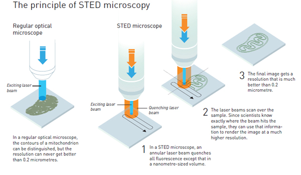
The principle of STED-microscopy
Article Stefan Helle, published in 1994 in the journal Optics Letters , has attracted attention, and he was offered a job at the Institute for Biophysical Chemistry in the Max Planck Society in Göttingen, where he could realize his theoretical assumptions in practice. Scientist made it to the 1999 building STED-microscope. In 2000, he published a picture bacteria Escherichia coli with unprecedented resolution.
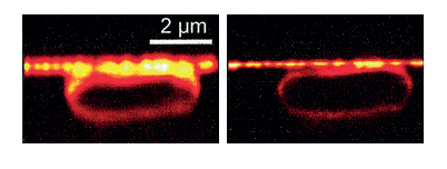
One of the first images taken by Stefan Helle. The bacterium Escherichia coli, the left – on the confocal microscope, the left – on the STED-microscope.
Another direction independently developed by American researchers Eric Bettsig and William Esko Merner. The latter with childhood name initials WE, to distinguish it from his father and grandfather, also William Merner. These scientists have developed methods to single-molecule fluorescence microscopy (single-molecule fluorescence microscopy).
Usually in the study of fluorescence, in particular the use of this phenomenon in microscopy, we deal with absorption and emission of radiation from a variety of molecules. In 1986, for the first time managed to Merner measure the absorption of light of a single molecule. Eight years later, Merner took the next step in their research by studying the green fluorescent protein ( green fluorescent protein , GFP, the role of this protein in modern biology we had once told). Merner found that the fluorescence of GFP in one embodiment can “turn on” and “off” as desired. When the protein absorbs light with a wavelength of 488 nanometers, the fluorescence starts but after some time will disappear. And, regardless of the amount of light directed to the protein, the response of the radiation occurs. However, if you use the light with a wavelength of 405 nm, the protein molecule fluoresces again.
Merner distributed GFT these molecules in the gel so that the distance between each individual molecule was larger than the Abbe diffraction limit. Due to the fact that they were scattered so rarely microscope was able to detect luminescence of individual molecules. About his work Merner told the magazine Nature in 1997.
The obtained results have helped Eric Merner Bettsigu, who worked in the field of near-field optical microscopy (near-field microscopy). He guessed that if the microscope will record the emission of molecules with a certain wavelength, scattered in the sample closer to each other than 0.2 m (approximate value of the Abbe limit), then the position can be determined with high accuracy. If the sample to be molecules with different properties, for example, when giving a response fluorescence radiation of different wavelengths, it is possible to make a separate picture of the distribution of each “kind” of molecules, and then superimposing them on each other in the full image resolution to achieve greater than Abbe limit. Molecules are distinguishable, even if the distance between them will be only a few nanometers.
These ideas Bettsig expressed in the publication in the journal Optics Letters in 1995, but it seemed impossible to implement them, so it does not contain molecules with desired properties. Bettsig even for a little while left academic activities and began working in his father’s company Ann Arbor Machine Company , engaged in applied research engineering developments. He continued to follow the publications and finally learned about research GFP. Returning to work in the field of microscopy, by 2005 Bettsig, experimenting with different types of fluorescent proteins, has developed a method to overcome the Abbe limit. Only molecules do not emit light of different colors, as he assumed in 1990, and began to glow at different points in time. That was enough to get a set of images, one of which will develop – is ultra-high resolution. The method was called microscopy localized photoactivation (photoactivated localization microscopy).
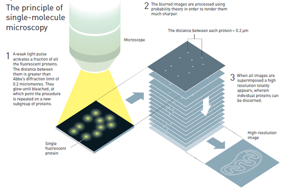
The principle of single-molecule microscopy (method localized photoactivation).
Now, thanks to single-molecule fluorescence spektoroskopii scientists can observe in real time the processes of protein folding and DNA and to determine the position of individual molecules in the cell to within a few tens of nanometers.
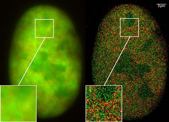
Image nucleus of cancer cells obtained by fluorescence microscopy, ultra-high resolution (right). With the help of green and red fluorescent proteins localized around 1200 molecules.
analogue of Helly, which uses two laser beams, has already found applications of the development of optical discs for recording high density information (on a standard disc fits 5 petabytes, which corresponds, for example, a video of about 10 years).
laureates themselves now used by their methods in the new study. Stefan Kjell looked inside living neurons to study at the molecular level structure of synapses in the brain. Merner research studies of proteins that play a role in the development of Huntington’s syndrome. Eric Bettsig explores the division of embryonic cells. After the work of Hella Merner and Bettsiga optical microscopy overcame Abbe diffraction limit and become a “nanoscopes».
No comments:
Post a Comment